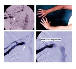
Affects 10-15% of all patients with TOS.
Venous TOS is characterized by subclavian vein compression between the clavicle and first rib. Most patients with venous TOS experience one or more of the following symptoms:
- Abrupt spontaneous swelling of the entire arm
- Cyanotic (bluish) discoloration of the arm
- Heaviness of the upper extremity
- Pain in the upper extremity
Axillary-subclavian vein “effort thrombosis” (also known as the “Paget-Schroetter” syndrome) is an unusual form of deep venous thrombosis (DVT) that occurs in young, active and otherwise healthy individuals, with no underlying blood clotting disorder. Thrombosis is the formation of blood clot inside of a blood vessel. The events or circumstances leading to effort thrombosis involve repetitive compression of the subclavian vein during activities involving arm elevation or exertion.
The initial phase of venous TOS is usually asymptomatic, involving compression injury of the vein between the clavicle and first rib, scar tissue formation, contraction around the outside of the vein, and fibrosis and wall thickening within the wall of the vein itself.
A blood clot does eventually form in the narrowed subclavian vein, leading to thrombotic occlusion. Growth and extension of the clot down the subclavian and into the axillary vein can then result in further obstruction of critical collateral veins, resulting in sudden symptoms that include substantial arm swelling and cyanotic (bluish) discoloration. Pulmonary embolism may also occur, particularly with motion of the arm, but this is infrequent compared to deep vein thrombosis, or DVT, in the lower extremities.
The diagnosis of venous TOS is based on the history and the symptoms present during clinical evaluation. Imaging studies, such as magnetic resonance angiography or catheter-based venograms, provide more definitive information on the location and extent of axillary-subclavian vein thrombosis than venous Duplex (ultrasound) studies. These studies can also be performed with the arms at rest and in elevation to provide additional information on the effects of arm position on the subclavian vein. Blood coagulation testing is often performed in patients with upper extremity DVT, but these tests are usually negative and add little to the initial diagnosis or management of effort thrombosis.
Conservative treatment of subclavian vein effort thrombosis has traditionally consisted of chronic anticoagulation (anti-clotting) therapies, intermittent arm elevation, long-term restrictions in arm activity, and the use of compression sleeves. Unlike lower extremity DVT, the proper duration of anticoagulation treatment for subclavian vein effort thrombosis is not known.
Because this condition is caused by repetitive mechanical compression of the vein rather than a disorder of blood clotting, in the absence of an alteration in the anatomy many physicians recommend lifelong anticoagulation.
To avoid further complications (such as recurrent thrombosis) and the need for long-term anticoagulation, current approaches to venous TOS emphasize the following:
- Early diagnosis by contrast venography
- Prompt initial use of catheter-based thrombolytic therapy to reduce the amount of thrombus within the axillary and subclavian veins
- Venous TOS surgery
In some cases, balloon angioplasty may be used at the same time as thrombolytic therapy in attempting to reduce the degree of stenosis in the subclavian vein. However, because the vein is usually obstructed by scar tissue in the wall of the vein as well as external compression between the clavicle and first rib, balloon angioplasty is often unsuccessful or results in only short-lived improvement. It has also become clear over the past decade that placement of stents within the subclavian vein frequently leads to poor outcomes and should be avoided.
In most patients, thrombolytic therapy is followed by immediate treatment with anticoagulation medications (heparin) and, after a variable interval, by surgical thoracic outlet decompression.
Venous TOS surgery is based on resection of the first rib. Robert Thompson, MD, TOS surgeon and director of the Washington University Center for Thoracic Outlet Syndrome at Barnes-Jewish Hospital, accompanies this with either endovascular management (balloon angioplasty) or direct venous reconstruction (patch angioplasty or bypass) for any residual subclavian vein stenosis.
Although excellent results have been reported for all of these surgical approaches, the best published outcomes have consistently been with approaches that involve direct reconstruction of the subclavian vein at the time of decompression surgery.
Venous TOS patients undergoing successful surgery are typically able to return to symptom-free normal activities within several months, including competitive athletics, without the need for long-term anticoagulation.
Unresolved issues regarding venous TOS are related to:
- Improving early detection and referral to a specialist for treatment
- Timing and methods used for thrombolysis
- The type and extent of surgical treatment needed to achieve optimal, long-term functional outcomes
- The occasional need for accompanying treatments, such as balloon angioplasty and chronic anticoagulation